Fdp And Fds Tendons : FDS-5 variations. Muscle: A. Normal, B. Absent, C. Unusual, D.... | Download Scientific Diagram. For many years, considerable conjecture has been expanded on the relationship between the fds and the fdp tendons and the role of the bifurcation and coming together of the fds around the fdp. Fds fdp fpl lumbricals origin from radial side of fdp. It is not enough just to observe the child make a fist as tendon injuries can be easily overlooked or missed. To check fds function, hold all adjacent fingers in extension and then release the finger you want to assess. · a nerve injury with a tendon injury may require.
The region locates between the fds insertion to distal palmar crease. The reconstruction of chronic flexor tendon injuries remains one of the more challenging injuries facing the hand and upper extremity surgeon. Of course, the smaller the holds are, and the studies have shown stress through the tendon pulley system and fdp is greater than the fds during crimp gripping, and hence it is more commonly injured. Flexor tendons and muscles these pictures of this page are about:fds and fdp tendons. · discontinuation of narcotics is expected, continue with tylenol and ibuprofen as needed.
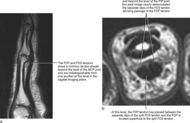
Flexor tendon injury and reconstruction | Plastic Surgery Key from plasticsurgerykey.com The fds tendons lie on the palmar side of the fdp tendons until they enter the a1 entrance of the digital sheath. The fdp passes through the tendinous hiatus just proximal to the tendinous chiasm (asterisk)and continues distally to insert on the distal phalanx. In this zone the fdp and fds flexor tendons enter the same fibrous sheath. This area is known as, no man's land, as historically results of tendon repair in this. Zone is unique in that fdp and fds in same tendon sheath (both can be injured within the flexor retinaculum). Each finger receives attachment from a fds and fdp tendon. The fds tendon tendons travel through the carpal tunnel, along with the fdp tendons, fpl tendon and median nerve. · a nerve injury with a tendon injury may require.
If a patient develops a contracture, they will sometimes need dynamic splinting about.
Adherence to the therapy program is advised to prevent a tendon rupture. The reconstruction of chronic flexor tendon injuries remains one of the more challenging injuries facing the hand and upper extremity surgeon. Both fdp and fds tendons in the digits receive dual nutritional supply from vascular perfusion and synovial diffusion 7 . The four fdp tendons course deep to the fds tendons through the flexor retinaculum and the carpal tunnel. The region locates between the fds insertion to distal palmar crease. This can be injured in an fdp avulsion injury. For many years, considerable conjecture has been expanded on the relationship between the fds and the fdp tendons and the role of the bifurcation and coming together of the fds around the fdp. Bakker at week 2 to have your stitches removed. Further, both fdp and fds tendons share the same. Ulna & interosseous membrane fdp: The fds and the fdp enter the fibrous sheath just proximal to the metacarpophalangeal joints. The fds tendon tendons travel through the carpal tunnel, along with the fdp tendons, fpl tendon and median nerve. Zone is unique in that fdp and fds in same tendon sheath (both can be injured within the flexor retinaculum).
In the setting of an intact flexor digitorum superficialis (fds), there are few indications for isolated flexor digitorum profundus (fdp) reconstruction. The flexor tendon system of the hand consists of the flexor muscles of the forearm, their tendinous extensions, and the specialized digital flexor sheaths. The flexor tendons of the fingers are the flexor digitorum superficialis (fds) and the flexor digitorum profundus (fdp). The reconstruction of chronic flexor tendon injuries remains one of the more challenging injuries facing the hand and upper extremity surgeon. The fds and fdp tendons should be tested individually;
IMAGING OF THE HAND AND FINGERS | Radiology Key from radiologykey.com Fds fdp fpl lumbricals origin from radial side of fdp. The fds and fdp tendons should be tested individually; Flexor tendons and muscles these pictures of this page are about:fds and fdp tendons. 5.zone v= fdp and fds tendons from the proximal edge of the transverse carpal ligament up to the volar forearm. Adherence to the therapy program is advised to prevent a tendon rupture. In this zone the fdp and fds flexor tendons enter the same fibrous sheath. In the setting of an intact flexor digitorum superficialis (fds), there are few indications for isolated flexor digitorum profundus (fdp) reconstruction. Disrupted, fds intact • two staged flexor tendon reconstruction • thumb flexor tendon reconstruction • secondary reconstruction in zones 3,4,5 • flexor tendon reconstruction in children • complications.
Angular joint displacement as a function of a controlled tendon excursion driven by computerized motors was used to quantify the kinematics of the finger.
· discontinuation of narcotics is expected, continue with tylenol and ibuprofen as needed. 5.zone v= fdp and fds tendons from the proximal edge of the transverse carpal ligament up to the volar forearm. This can be injured in an fdp avulsion injury. To check fds function, hold all adjacent fingers in extension and then release the finger you want to assess. Zone is unique in that fdp and fds in same tendon sheath (both can be injured within the flexor retinaculum). Of course, the smaller the holds are, and the studies have shown stress through the tendon pulley system and fdp is greater than the fds during crimp gripping, and hence it is more commonly injured. It is not enough just to observe the child make a fist as tendon injuries can be easily overlooked or missed. The flexor tendon system of the hand consists of the flexor muscles of the forearm, their tendinous extensions, and the specialized digital flexor sheaths. This area is known as, no man's land, as historically results of tendon repair in this. • tenolysis • acute free tendon graft • single stage flexor tendon grafting with fdp. The reconstruction of chronic flexor tendon injuries remains one of the more challenging injuries facing the hand and upper extremity surgeon. 5 fdp simultaneous flexion of multiple digits origin: Further, both fdp and fds tendons share the same.
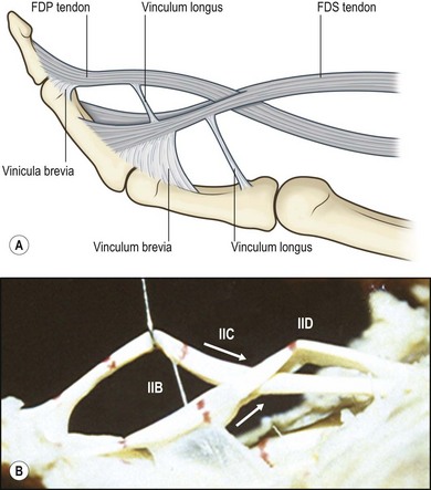
Common muscle origin for several tendons simultaneous 8 camper's chiasma fds divides and passes around the fdp tendon, the two portions of the fds reunite at camper's chiasma. The four fdp tendons course deep to the fds tendons through the flexor retinaculum and the carpal tunnel. The fdp passes through the tendinous hiatus just proximal to the tendinous chiasm (asterisk)and continues distally to insert on the distal phalanx. The fds tendon graft is sutured to the prosthetic graft; This can be injured in an fdp avulsion injury.
Flexor Tendon Injuries - Hand - Orthobullets from upload.orthobullets.com To check fds function, hold all adjacent fingers in extension and then release the finger you want to assess. The reconstruction of chronic flexor tendon injuries remains one of the more challenging injuries facing the hand and upper extremity surgeon. The fds tendons split at camper's chiasm to allow fdp tendon to pass through. Disrupted, fds intact • two staged flexor tendon reconstruction • thumb flexor tendon reconstruction • secondary reconstruction in zones 3,4,5 • flexor tendon reconstruction in children • complications. These components work in concert to produce smooth and efficient flexion of the individual digits of the hand. Fds splints so fdp can go through. Zone is unique in that fdp and fds in same tendon sheath (both can be injured within the flexor retinaculum). Both fdp and fds tendons in the digits receive dual nutritional supply from vascular perfusion and synovial diffusion 7 .
These components work in concert to produce smooth and efficient flexion of the individual digits of the hand.
The reconstruction of chronic flexor tendon injuries remains one of the more challenging injuries facing the hand and upper extremity surgeon. Further, both fdp and fds tendons share the same. All flexor tendon approaches protect the flexor tendon repair with some version of a dorsal blocking orthosis that flexes the wrist and/or mp joints zone ii: Each finger receives attachment from a fds and fdp tendon. The fdp and fpl tendons are found in the deepest level of the carpal tunnel. · discontinuation of narcotics is expected, continue with tylenol and ibuprofen as needed. Adherence to the therapy program is advised to prevent a tendon rupture. Fds and fdp tendons (page 1) flexor tendon laceration the fds tendons to any single finger may be tested by holding the other. Vascular perfusion is provided by the fdp and both vincula rupture with no fractures tendon retracts into palm, presenting as a tender lump early operative repair necessary. For many years, considerable conjecture has been expanded on the relationship between the fds and the fdp tendons and the role of the bifurcation and coming together of the fds around the fdp. Zone is unique in that fdp and fds in same tendon sheath (both can be injured within the flexor retinaculum). If a patient develops a contracture, they will sometimes need dynamic splinting about. Both fdp and fds tendons in the digits receive dual nutritional supply from vascular perfusion and synovial diffusion 7 .
In the setting of an intact flexor digitorum superficialis (fds), there are few indications for isolated flexor digitorum profundus (fdp) reconstruction fdp. Common muscle origin for several tendons simultaneous 8 camper's chiasma fds divides and passes around the fdp tendon, the two portions of the fds reunite at camper's chiasma.
Share :
Post a Comment
for "Fdp And Fds Tendons : FDS-5 variations. Muscle: A. Normal, B. Absent, C. Unusual, D.... | Download Scientific Diagram"


Post a Comment for "Fdp And Fds Tendons : FDS-5 variations. Muscle: A. Normal, B. Absent, C. Unusual, D.... | Download Scientific Diagram"